About the lab
The Beckstein Lab is part of the Center for Biological Physics and the Department of Physics at Arizona State University where it has been active since 2012 under the direction of Oliver Beckstein.
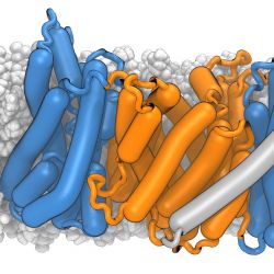
We use computational approaches to explore how biomolecules function at the molecular level, with a focus on membrane proteins. Our research lies at the intersection of physics, biology, and computation, aiming to predict protein function and activity from structure alone and to reveal the molecular principles underlying biological processes. In particular, we study transmembrane transport mechanisms mediated by membrane proteins such as secondary active transporters and ion channels. This work contributes to fields including structural biology, physiology, nanobiotechnology, and drug discovery.
The lab also develops open-source software for the molecular sciences, emphasizing robust and widely usable tools for data analysis. We are part of the leadership team of the MDAnalysis Project , creators of the MDAnalysis Python package—one of the most widely used tools for analyzing particle-based molecular simulations.